Ts Of Compact Bone Diagram - Page 2 Compact Bone High Resolution Stock Photography And Images Alamy : Flat bones, like those of the cranium, consist of a layer of diploë (spongy bone), lined on either side by a layer of compact bone ().
Ts Of Compact Bone Diagram - Page 2 Compact Bone High Resolution Stock Photography And Images Alamy : Flat bones, like those of the cranium, consist of a layer of diploë (spongy bone), lined on either side by a layer of compact bone ().. There are two types of bone tissue: The functional units of compact bone are osteons; Which contain a centrally located haversian canal, encased in lamellae (concentric rings). It can be found under the periosteum and in the diaphyses of long bones, where it provides support and protection. Learn with flashcards, games, and more — for free.
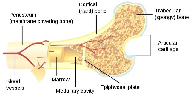
Some, mostly older, compact bone is remodelled to form these haversian systems (or osteons). Diagram of a typical long bone showing both compact (cortical) and cancellous (spongy) bone. Because of its strength, the compact bone makes it possible for the bone to support weight. The hollow region in the diaphysis is called the medullary cavity, which is filled with yellow marrow. Diagram of haversian canal.an osteon comprises a long, hollow central canal that is surrounded by concentric layers called lamallae.
2 Schematic Depiction Of Compact Bone Structure 12 Download Scientific Diagram from www.researchgate.net Compact bone, also called cortical bone, dense bone in which the bony matrix is solidly filled with organic ground substance and inorganic salts, leaving only tiny spaces (lacunae) that contain the osteocytes, or bone cells.compact bone makes up 80 percent of the human skeleton; It makes up the outer cortex of all bones and is in immediate contact with the periosteum. Bone marrow cavity _ b. Compact bone, also called cortical bone, is the hard, stiff, smooth, thin, white bone tissue that surrounds all bones in the human body. Anatomy of a long bone. The remainder is cancellous bone, which has a spongelike appearance with numerous large spaces and is found in the. Which contain a centrally located haversian canal, encased in lamellae (concentric rings). Ʒ ən / (named for clopton havers ) is the fundamental functional unit of much compact bone.
Label the components of compact bone on the following diagram.
Compact bone, also called cortical bone, dense bone in which the bony matrix is solidly filled with organic ground substance and inorganic salts, leaving only tiny spaces (lacunae) that contain the osteocytes, or bone cells.compact bone makes up 80 percent of the human skeleton; This type of bone is located between layers of compact bone and is thin and porous. There are small canals that run through the bone, which allow blood vessels to penetrate it. Compact bone is formed in concentric circles. Flat bones, like those of the cranium, consist of a layer of diploë (spongy bone), lined on either side by a layer of compact bone (). This is the area of bone to which ligaments and tendons attach. Compact and spongy.the names imply that the two types differ in density, or how tightly the tissue is packed together. As seen in the image below, compact bone forms the cortex, or hard outer shell of most bones in the body. The remaining surface of each bone is covered with a thin connective tissue membrane called the periosteum, which contains numerous blood vessels, nerves, and lymphatic vessels. The cells of compact bone, which is also called cortical bone, appear to be tightly packed into a solid mass. Because of its strength, the compact bone makes it possible for the bone to support weight. Cartilage to prevent bone from rubbing directly on bone. It makes up the outer cortex of all bones and is in immediate contact with the periosteum.
This central canal is referred to as the haversian canal. (b) in this micrograph of the osteon, you can clearly see the concentric lamellae and central canals. It can be found under the periosteum and in the diaphyses of long bones, where it provides support and protection. The diaphysis is the tubular shaft that runs between the proximal and distal ends of the bone. The remaining surface of each bone is covered with a thin connective tissue membrane called the periosteum, which contains numerous blood vessels, nerves, and lymphatic vessels.
Compact Bone Definition And Function Biology Dictionary from biologydictionary.net Compact bone, also called cortical bone, dense bone in which the bony matrix is solidly filled with organic ground substance and inorganic salts, leaving only tiny spaces (lacunae) that contain the osteocytes, or bone cells.compact bone makes up 80 percent of the human skeleton; In long bones, as you move from the outer cortical compact bone to the inner medullary cavity, the bone transitions to spongy bone. Although the calls are close together, this type of bone is not completely solid. Compact bone, also called cortical bone, is the hard, stiff, smooth, thin, white bone tissue that surrounds all bones in the human body. 13 photos of the compact bone diagram labeled. Flat bones, like those of the cranium, consist of a layer of diploë (spongy bone), lined on either side by a layer of compact bone (). This type of bone is located between layers of compact bone and is thin and porous. This is the area of bone to which ligaments and tendons attach.
The walls of the diaphysis are composed of dense and hard compact bone.
Bone marrow diagram, compact bone diagram quiz, compact bone slide labeled, diagram long bone, labeled compact bone model, human anatomy, bone marrow diagram, compact bone diagram quiz, compact bone slide labeled, diagram long bone, labeled compact bone model. Diagram of a typical long bone showing both compact (cortical) and cancellous (spongy) bone. Compact bone is formed in concentric circles. Diagram of haversian canal.an osteon comprises a long, hollow central canal that is surrounded by concentric layers called lamallae. The walls of the diaphysis are composed of dense and hard compact bone. Osteoblasts on the inside of the periosteum deposit compact bone over the spongy bone. If the outer layer of a cranial bone fractures, the brain is still protected by the intact inner layer. This central canal is referred to as the haversian canal. The functional units of compact bone are osteons; This type of bone is located between layers of compact bone and is thin and porous. It is dense (because of calcified matrix) with tiny spaces known as lucanas. Compact bone is the denser, stronger of the two types of bone tissue ( (figure) ). Because of its strength, the compact bone makes it possible for the bone to support weight.
Bone marrow cavity _ b. Flat bones, like those of the cranium, consist of a layer of diploë (spongy bone), lined on either side by a layer of compact bone (). Concentric lamellae interstitial lamellae central canal lacuna osteocyte canaliculus. It is also called osseous tissue or cortical bone and it provides structure and support for an organism as part of its skeleton, in addition to being a location for the storage of minerals like calcium.about 80% of the weight of the human skeleton comes from. (b) in this micrograph of the osteon, you can clearly see the concentric lamellae and central canals.
Compact Bone Definition Structure Function Video Lesson Transcript Study Com from study.com Anatomy of a long bone proximal epiphysis diaphysis distal epiphysis compact bone spongy bone medullary cavity. (b) in this micrograph of the osteon, you can clearly see the concentric lamellae and central canals. The functional units of compact bone are osteons; This is the area of bone to which ligaments and tendons attach. Ʒ ən / (named for clopton havers ) is the fundamental functional unit of much compact bone. You can think of compact bone as being very similar. Exercise 7.2 spongy bone 2 which of the following is/are visible on a slide of decalcified spongy (cancellous) bone a. Human compact bone is composed of parallel columns made up of concentric bony layers called lamellae organized around channels containing blood vessels, lymph vessels and nerves.
(b) in this micrograph of the osteon, you can clearly see the concentric lamellae and central canals.
Diagram of a typical long bone showing both compact (cortical) and cancellous (spongy) bone. Although the calls are close together, this type of bone is not completely solid. Compact bone, or cortical bone, mainly serves a mechanical function. Compact bone is formed in concentric circles. If the outer layer of a cranial bone fractures, the brain is still protected by the intact inner layer. About press copyright contact us creators advertise developers terms privacy policy & safety how youtube works test new features press copyright contact us creators. 13 photos of the compact bone diagram labeled. (b) in this micrograph of the osteon, you can clearly see the concentric lamellae and central canals. Exercise 7.2 spongy bone 2 which of the following is/are visible on a slide of decalcified spongy (cancellous) bone a. The diaphysis is the tubular shaft that runs between the proximal and distal ends of the bone. Human compact bone is composed of parallel columns made up of concentric bony layers called lamellae organized around channels containing blood vessels, lymph vessels and nerves. In long bones, as you move from the outer cortical compact bone to the inner medullary cavity, the bone transitions to spongy bone. It can be found under the periosteum and in the diaphyses of long bones, where it provides support and protection.
Compact bone stands in stark contrast to trabecular bone in several ways compact bone diagram. The dense and hard exterior surface bone is called cortical or compact bone.


0 Komentar